
Foot & Ankle
Foot & Ankle Anatomy
What is the Normal Anatomy of the Foot and Ankle?
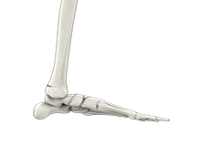
The foot and ankle are a complex joint involved in movement and providing stability and balance to the body. The foot and ankle consist of 26 bones, 33 joints, and many muscles, tendons and ligaments.
Bones of the Ankle
The ankle joint connects the leg with the foot and is composed of three bones: tibia, fibula and talus. The tibia or shinbone and fibula or calf bone are bones of the lower leg which articulate with the talus or ankle bone, enabling up and down movement of the foot.
Three bony bumps present on the ends of the tibia and fibula form parts of the ankle joint:
- The medial malleolus, formed by the tibia, is found on the inside of the ankle.
- Posterior malleolus, also formed by the tibia, is found at the back of the ankle.
- Lateral malleolus, formed by the fibula, is found on the outer aspect of the ankle.
Bones of the Feet
The foot acts as a single functional unit, but can be divided into three parts: the hindfoot, midfoot and forefoot.
The hindfoot forms the ankle and heel and is made up of the talus bone and calcaneus or heel bone. The heel bone is the largest bone in the foot.
The midfoot connects the hindfoot to the forefoot, and consists of one navicular bone, one cuboid bone, and three cuneiform bones. The navicular bone is found in front of the heel bone, and the cuneiform and cuboid bones are arranged in front of the navicular bone.
These bones are connected to five metatarsal bones of the forefoot, which form the arch of the foot for shock absorption while walking or running. The forefoot is also made up of the toes or digits, formed by phalanges, three in each toe, except the big toe, which has only two phalanges. The big toe has two additional tiny round sesamoid bones in the ball of the foot, which help in upward and downward movement of the toe.
Ankle and Foot Joints
There are 33 joints in the ankle and foot. They include:
- Hinge joints in the ankle, which allow flexion (bending) and extension
- Gliding joints found in the hindfoot, which allow gliding movements
- Condyloid joints found in the forefoot and toes, which allow the flexion (bending) and extension, adduction and abduction (sideward movement).
The joints of the foot and ankle provide stability and support the weight of the body, helping you to walk or run, and to adapt to uneven ground.
The joint surface of all bones of the ankle and foot are lined by a thin, tough, flexible, and slippery surface called articular cartilage, which acts as a shock absorber and cushion to reduce friction between the bones. The cartilage is lubricated by synovial fluid, which further enables smooth movement of the bones.
Soft Tissues of the Ankle and Foot
Our feet and ankle bones are held in place and supported by various soft tissues such as cartilage, ligaments, muscles, tendons and bursae.
Cartilage is the flexible, shiny, smooth tissue on the ends of bones that meet to form a joint. Cartilage provides cushioning between the bones allowing smooth movement.
Ligaments are tough rope-like tissue that connect bones to other bones, and holds them in place providing stability to the joints. The plantar fascia is the largest ligament in the foot, originating from the heel bone to the forefoot, it extends along the bottom surface of the foot and is involved in maintaining the arch of the foot. The plantar fascia ligament stretches and contracts to provide balance and strength to the foot. Lateral ligaments on the outside of the foot and medial ligaments on the inside of the foot provide stability and allow up and down movement of the foot.
The foot is made up of 20 muscles, which help in movement. The main muscles include:
- Anterior tibial muscle: allows up and down movement of the foot
- Posterior tibial muscle: supports the arch
- Peroneal tibial muscle: controls movement on the outside of the ankle
- Extensors: enable the ankle to raise the toes just before stepping forward
- Flexors: stabilize the toes against the floor
- Smaller muscles are also present to help the toes lift and curl.
Tendons are soft tissues that connect muscles to bones. The largest and strongest tendon in the foot is the Achilles tendon, present at the back of the lower leg around the heel bone. Other tendons include peroneal and anterior and posterior tibialis.
Bursae
Bursae are small fluid-filled sacs that decrease friction between tendons and bone or skin. Bursae contain special cells called synovial cells that secrete a lubricating fluid.
Ankle Fracture
The ankle joint is composed of three bones: the tibia, fibula and talus, which are articulated together. The ends of the fibula and tibia (lower leg bones) form the inner and outer malleolus, which are the bony protrusions of the ankle joint that you can feel and see on either side of the ankle. The joint is protected by a fibrous membrane called a joint capsule, and filled with synovial fluid to enable smooth movement.
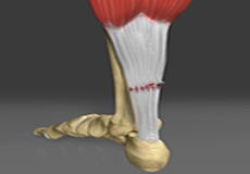
Achilles Tendon Rupture
The Achilles tendon is a strong, fibrous cord present behind the ankle that connects the calf muscles to the heel bone. It is used when you walk, run and jump. The Achilles tendon ruptures most often in athletes participating in sports that involve running, pivoting and jumping. Recreational sports that may cause Achilles rupture include tennis, football, basketball and gymnastics.
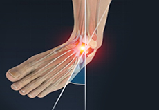
Ankle Sprain
A sprain is the stretching or tearing of ligaments, which connect adjacent bones and provide stability to a joint. An ankle sprain is a common injury that occurs when you suddenly fall or twist the ankle joint or when you land your foot in an awkward position after a jump. Most commonly it occurs when you participate in sports or when you jump or run on a surface that is irregular.

Common Toe Deformities
Toes are the digits in your foot and are associated with walking, providing balance, weight-bearing and other activities. A variety of toe deformities occur in children's feet.
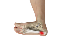
Plantar Fasciitis
Plantar fasciitis refers to inflammation of the plantar fascia, a thick band of tissue that is present at the bottom of the foot. It runs from the heel bone to the toe and forms the arch of your foot. Plantar fasciitis is one of the most common causes of heel pain. It is most often seen in middle-aged men and women, but may also occur in those who are constantly on their feet.
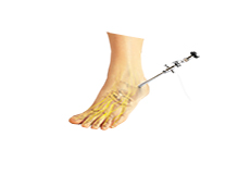
Ankle Arthroscopy
Ankle arthroscopy is a minimally invasive surgical procedure in which an arthroscope, a small, soft, flexible tube with a light and video camera at the end, is inserted into the ankle joint to evaluate and treat a variety of conditions.
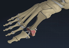
Bunion Surgery
A bunion, also known as hallux valgus, is bony prominence at the base of the big toe, which often results in pain, redness and rubbing in footwear. The first metatarsal bone abnormally angles outward towards the other foot from its joint in the midfoot. A bunion can change the shape of your foot, make it difficult for you to find shoes that fit correctly and worsen the symptoms if left untreated.